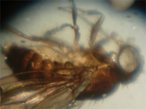
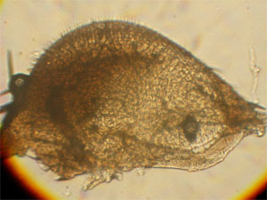
Microscope 124. Vignetting, chromatic aberration, low magnification, difficult focusing. Note poor resolution, espec eye bristles.
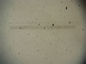
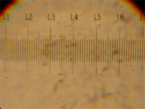
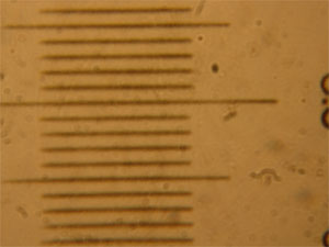
Scalebar is 2mm across. Image is appx 28x
Microscope 18. Chromatic aberration, difficult focusing, low magnification. Appx 78x. Resolution is better, but you cannot see bristles between eye lenses, only around the compound eye. This is reflected light because the scope has poor transmitted light. Axial alignment is extremely poor, so light from substage mirror misses the objective.
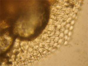
Dissected compound eye. Microscope 167, using two objective lenses (00 + 1). Chromatic aberration. Much easier focusing (coarse only), better lighting because of the primative condenser and much better axial alignment. Magnification appx 116x. Note how we can just make out the bristles.
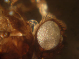
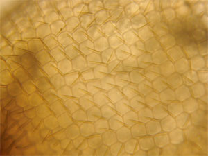
Microscope 188. No chromatic aberration, coarse and fine focus, very good condenser for even illumination, high magnification. Mag appx 350x. The bristles between lenses can be resolved.
Example images from four historical microscopes.
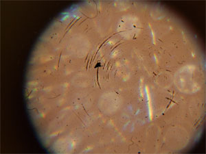
This is a picture of diatoms taken through scope 276. Look at the lens grinding marks found on the objective lens. Refraction through scratches from the circular grinding motion produce the black arcs you see here. Note also the poor imaging of the diatoms. Aberrations like this (chromatic, vignetting, low mag, light loss) were common in lenses of this era (pre 18thC). Scratches, bubbles, soot, etc are the cause of this poor lens quality.
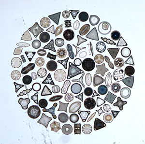
Right is a low mag (2.5x) pix of the diatom circle using a modern compound microscope.