 |
|||||
 |
 |
||||
 |
|||||
 |
 |
||||

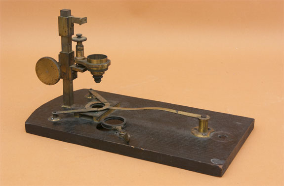
This is part of a device designed and made by Alfred Donné (1801–1878) and Léon Foucault (1819–1868) that was used to make the first published medical photomicrographs. This device was a projecting microscope attached to an arc lamp (the source of Foucault's name: "photo-électrique") placed inside a large enclosure. It functioned to project a magnified image horizontally, much like the Solar Microscope and Magic Lantern of the era. The Daguerre technique (invented by L.J.M. Daguerre (1789–1851) was used to make a "daguerrotype" photograph. Examples of the Donné/Foucault daguerrotypes are found here.
The instrument consists of a square support pillar mounted to a rectangular, black-painted wooden base. There is one stacked objective, with two lenses ("1" & "2"). The objective is mounted to a cantilever, as is the spring-loaded, threaded fine focus mechanism. Coarse focus is typical rack & pinion. Above the objective is the mount for a projection lens, which was used to project the image onto a wall or daguerrotype plate. Samples (slide preparations) were fixed to the "stage" (a metal plate with a hole) using either two stage clips or a circular spring mechanism. Adjacent to the sample stage Foucault drilled a slanted observation hole with a nearly opaque filter. This was used presumably to adjust the illuminating electrodes (which eroded rapidly). The heat generated would have been substantial, and this is witnessed by the charred base on the reverse side. This instrument in the Golub Collection is the only component remaining from Léon Foucault's complete device. It was No. 84 in the original Nachet Collection of Paris.
Included with the Foucault microscope is an optical device engraved "The "DAVON" (Regd) Micro-Telescope". This device cound not have been used by Foucault as it was invented well after Foucault's device. It is unclear why the Davon was included with the Foucault device.
For a thorough description of Foucault's Photo-électrique device see
Tobin, W. 2006. Alfred Donné and Léon Foucault: The first applications of electricity and photography to medical illustration. J. Vis. Com. Med 29(1): 6-13.
1886 thesis by Jean-Georges Bernard on the ‘History of the microscopes and what medicine owes to them’ (thanks to Jeroen Meeusen)