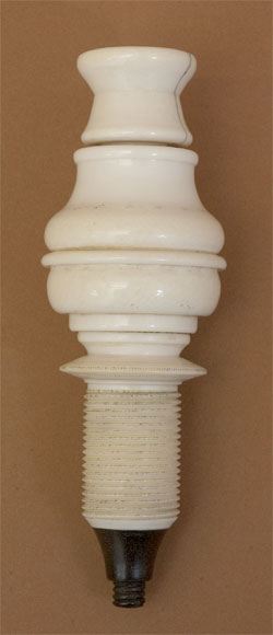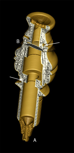
3D model of microscope No. 253 rendered from a CT Scan series shows only two imaging lenses exist in this instrument: an objective lens (A) and the eyelens (B). Both lenses are biconvex. The two seams (arrows) in the body of the microscope suggests that the instrument was made from three separate pieces of ivory: threaded stem, central body, and top eyecup. The top eyepiece insert and objective nosepiece are made from a different material; probably the hardwood Lignum vitae.
The construction of the microscope and the placement and number of imaging lenses suggests that this instrument was made before the introduction of the Field lens by Monconys in 1660 and adopted by Campani in 1661.
The microscope was imaged by Dr. Gary Richards of the University of the Pacific, San Francisco using a medical CT Scanner. This 3D model was created from the DICOM image series using OsiriX 3.8.